Considering that nearly 40 million magnetic resonance imaging (MRI) scans are performed in the United States each year, there’s a reasonably good chance that you’ll have this test at some point in your lifetime (if you haven’t already). MRIs are so commonly used because they’re a noninvasive way to visualize much of the inside of our bodies.
MRI is relatively safe (see below for possible risks), but as with any diagnostic test, having one can be an anxiety-producing experience—especially if you have claustrophobia, since the closed version is a tunnel-shaped machine that you slide into. Going into the test unaware could increase the stress. About 20 percent of people surveyed for a 2024 study in Scientific Reports said their referring doctor didn’t tell them what to expect during an MRI, or offered little information about it, which affected their perception of the test.
If you’re scheduled to have your first MRI, you’ll feel more comfortable going in if you understand how this scan works, what conditions it can detect, and what to expect.
MRI basics
MRI doesn’t use ionizing radiation to take images of inside your body. (This is a big plus over X-rays and CT scans, which do use radiation.) Instead, it uses powerful magnets. The magnetic field it produces temporarily changes the position of positively charged particles called protons in your body, causing them to release signals that the machine detects. A computer converts those signals into an image of whatever body part is being scanned.
Traditional MRI is a closed tube. Open MRI is an alternative that’s open on both sides. It may be preferable for people with claustrophobia, or for those whose height or weight makes a closed MRI too uncomfortable, though the images may not be as clear as those from a closed MRI. With some open MRIs you sit or stand instead of lying down.
Peeking inside
Here are some reasons why your doctor might send you for an MRI:
- To diagnose a brain or spinal cord condition, such as an ischemic stroke (not a hemorrhagic stroke), tumor, injury, aneurysm, or multiple sclerosis
- To find blockages or inflammation in blood vessels
- To check for tumors or structural problems in organs like the liver, kidneys, pancreas, spleen, uterus, ovaries, or prostate
- To detect breast cancer, especially if you’re at high risk or you have dense breast tissue
- To diagnose soft tissue injuries, such as to a muscle
Functional MRI (fMRI) is a special type of MRI that detects blood flow to certain areas of your brain. Your doctor might use this test in the planning stage of surgery or radiation therapy to identify which parts of your brain handle certain tasks (such as writing or speaking), in order to avoid damaging them.
MRI isn’t the optimal imaging scan in certain situations. A CT scan (which combines X-ray images to show cross-sections of your organs and other tissues) might be preferable to an MRI if you’ve had a head injury (CT scans are better at detecting bleeding in the brain), you have a bone condition or injury, you have severe claustrophobia, or you are unable to lie still (CT scans are faster and less affected by motion). To detect a simple bone fracture, all you might need is a regular X-ray. If you have a metal-containing implant, depending on the type, an MRI might not be safe.
MRI offers clearer images of the brain, spinal cord, and soft tissues like muscles, ligaments, and tendons than do X-rays or CT scans. This makes MRI more useful for diagnosing brain conditions like tumors and aneurysms, as well as for knee and shoulder injuries involving tendons and ligaments. If you need frequent imaging tests, MRI may be the better choice because it doesn’t expose you to radiation like X-rays and CT scans do. However, it is usually more expensive for people without health insurance, and those who are insured may find that their carrier won’t cover MRI if a less costly option is available.
For a brighter look, add contrast
Usually injected into a vein before an MRI, contrast agents, such as the rare earth metal gadolinium, make your organs, blood vessels, and tissues show up brighter on the image.
Contrast is helpful for diagnosing conditions like cancer, multiple sclerosis, or a problem with blood flow in the brain or spinal cord, but it isn’t safe for everyone. Some people are allergic to the contrast agent. Those with kidney disease are at increased risk for a rare but life-threatening condition called nephrogenic systemic fibrosis (NSF)—a thickening of the skin, organs, and other tissues that’s been linked to MRI contrast agents. Your doctor might send you for kidney function blood tests before referring you for an MRI with contrast, especially if you’re over age 60 or you have kidney disease.
Getting ready
An MRI doesn’t require much preparation. You can usually eat, drink, and take your medications as normal (your doctor or the imaging center will let you know if you need to fast or stop any medicines). Before your visit, ask the referring doctor what they are trying to learn from the scan, and which part of your body will be visualized.
If you are having an MRI with contrast, let your doctor or the radiologic technologist know whether you’ve ever had an allergic reaction to the contrast material being used in the scan, and if you have kidney disease.
Also tell them if you have claustrophobia. You can request an open MRI when you schedule the test (while keeping in mind that it isn’t completely open and it may not be available at that facility or for the type of scan you are having). Your doctor may be able to prescribe a short-acting sedative before the test. You might bring a friend or family member with you for support, or to drive you home after the test if you receive sedation. Relaxation techniques such as deep breathing and meditation can help you stay calm during the test, and the MRI team will work with you to make sure you’re comfortable.
Because the MRI machine uses a powerful magnet, certain types of metal inside your body could be dangerous (the same goes for metals outside your body, which is why you’ll be told to remove all jewelry). Its magnetic field is about 21,000 times greater than the earth’s magnetic field, and it will pull in any metal objects at a speed of 40 mph. Certain older cerebrovascular clips are unsafe inside the machine, but most modern metallic implants (such as hip prostheses, mechanical heart valves, coronary stents, and sutures in the sternum) don’t attract magnets and pose no hazard. You will be asked if you have any metal in your body. The technologist may also run a metal-detecting wand over your body to check for sources of metal you might not know about.
Dress for the scan in loose-fitting, comfortable clothing that contains no metal snaps or zippers. For an upper or lower body scan, you might want to wear a two-piece outfit so that you can remove the top or bottom only. The technologist may give you scrubs or a hospital gown to wear during the scan. Leave any metal objects, such as jewelry, glasses, hearing aids, phone, dentures, hairpins, coins, and keys at home or in the locker provided at the imaging center. Don’t wear makeup, because some cosmetics may contain metal without your knowledge.
Time in the tube
For an MRI with contrast, a nurse or trained technician will first place an intravenous (IV) catheter into a vein in your arm or hand. The contrast agent will be administered through the IV at specific points during the scan.
You’ll lie on the table face up (for brain or abdominal scans) or face down (for breast and some back scans)—or you might be sitting or standing. For some scans, you’ll get an MRI coil—a frame that fits over and surrounds the body part being scanned (such as the head, knee, or breast). The coil acts like an antenna, helping the MRI machine get clearer images.
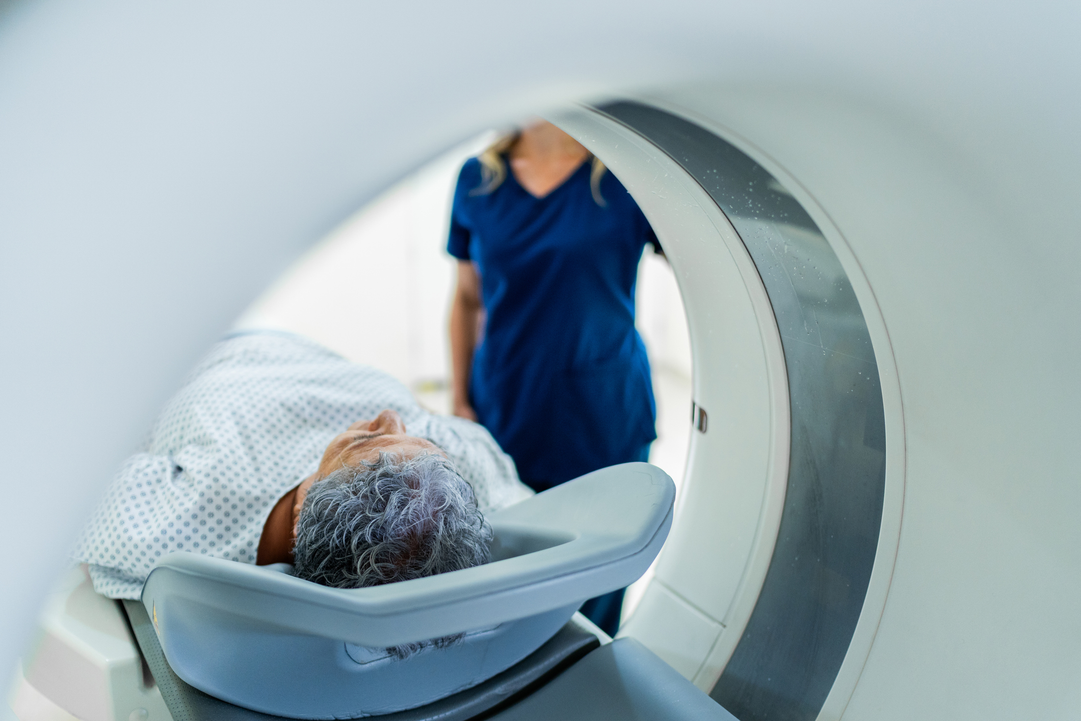
Once you’re ready, the technologist will slide you into the machine until the part of your body that’s being imaged is in the middle. You’ll be able to hear the technologist during the test, and you can use the alarm button they’ll place in your hand to alert them of any problems that occur while the scan is underway.
MRI scans are very noisy. You’ll hear loud, alternating buzzing, banging, and knocking. These sounds come from the gradient coils—looped wires surrounding the magnet that carry the currents used to produce the magnetic field. The technologist will give you earplugs or MRI-safe headphones (sometimes playing the music of your choice) to partially drown out the sounds.
You’ll need to lie as still as possible during the scan. Any movement could compromise the clarity of the images.
After the MRI
An MRI typically takes 30 to 50 minutes, depending on what part of your body is being scanned and the type of machine used. Afterward, you can go back to your usual activities, unless you took a sedative for the scan (in which case, it has to wear off before you can drive or be active).
There shouldn’t be any side effects, other than a little potential soreness at the injection site if contrast was used, and possibly nausea. People who have a reaction to the contrast agent can experience mild symptoms such as itching and hives. Only in rare cases does the contrast agent trigger a severe allergic reaction, with symptoms like a drop in blood pressure, difficulty breathing, or a serious heart problem.
A radiologist will analyze your MRI images and send the report to the doctor who ordered the scan. Your doctor should share results with you in a few business days. You’ll get the results in your patient portal (by law), possibly before your doctor calls you. Still, it’s best to wait for your doctor to share the results with you, since the findings might be very technical and difficult to understand on their own.
BOTTOM LINE: An MRI is a useful imaging test for diagnosing conditions like brain and spine disorders, soft tissue injuries, and cancer. Before you have the test, discuss with your doctor why it’s being done and what it’s expected to reveal. To avoid surprise bills, make sure your insurance company has preauthorized the test (most insurance plans require preauthorization, although it depends on your specific plan and how the test is coded), and ask how much you’ll have to pay out of pocket.